:max_bytes(150000):strip_icc()/GettyImages-308783-003-e6958f3f1e50487c93b25596348056cd.jpg)

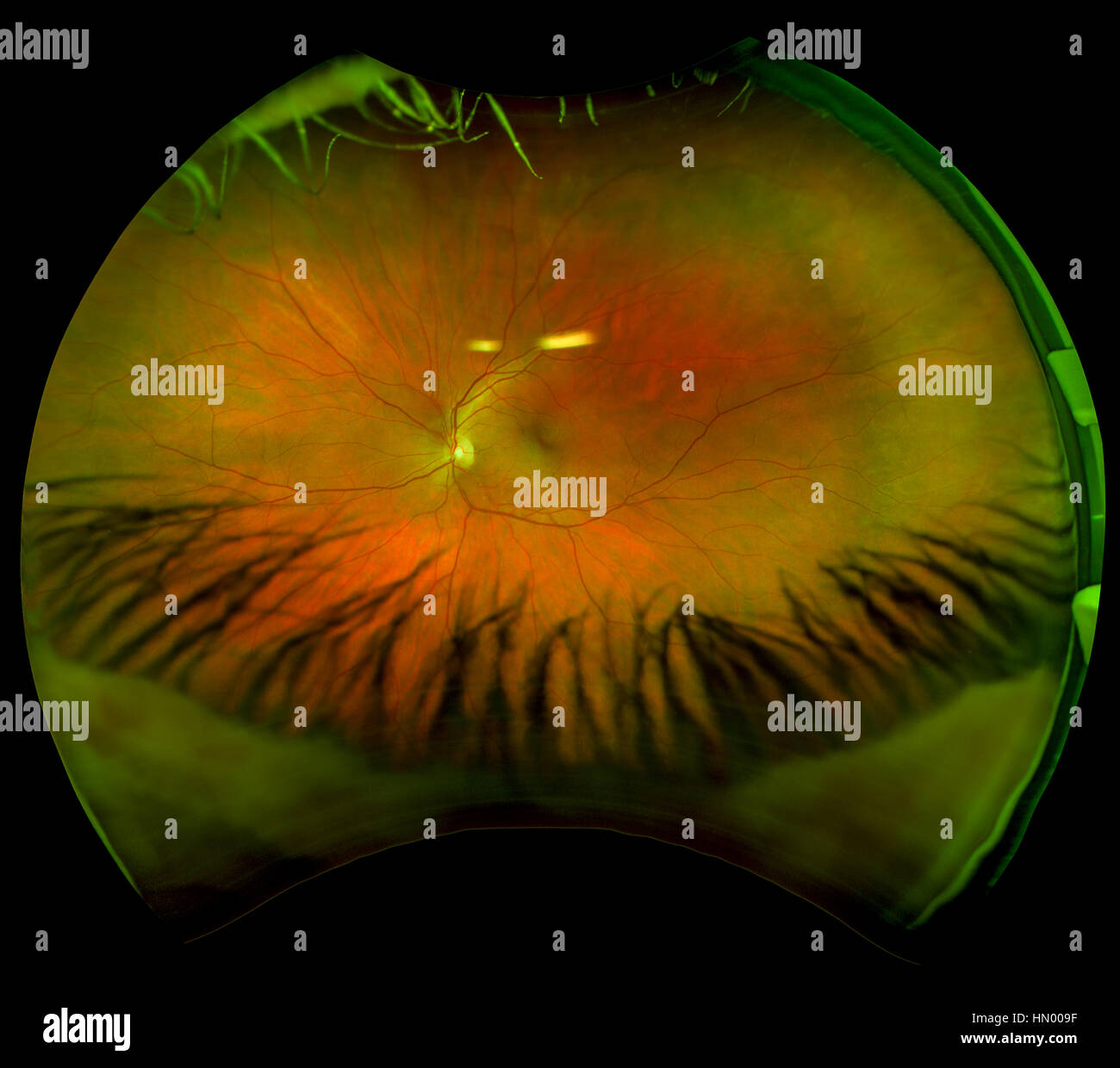
Glaucoma, which causes damage to the optic nerve.Vision-threatening conditions that can be detected with retinal imaging include: With these results we can refer the patient to an ophthalmologist for further tests and treatment. Many eye conditions can cause irreversible damage before any obvious symptoms are noticeable, but changes to the retina can indicate a problem in its early stages. By studying the images produced, our optometrist can search for signs of eye disease. We invested in the advanced optomap ultra-widefield camera to give our patients the most comprehensive eye examination. A high-resolution panoramic digital image is captured, covering 200° of the retina. We use an Optos Daytona ultra-widefield camera, which scans your eye using low-powered laser technology. The image of your retina produced is one of the best ways to check your eye health. Retinal imaging is suitable for patients of all ages and we recommend it as part of our advanced eye exam. Within seconds a digital image is taken and the results can be reviewed by the optometrist right away. Simply position your head using the chin and headrests, keep still and follow the optometrist’s directions about where to look. An optomap machine does all the work in just a moment.

The first fundus cameras were contraptions that had to be strapped to the head, but now it’s much easier to take a photo of your retina. As photography advanced, so did fundus technology, with a view increasing from just 15% to 82% of the retina which we can now capture in a single image. It took some time to develop the first cameras capable of this, and of course, back then they were very basic. Optometrists realised over a century ago that fundus photography, or photography of the back of the eye, could be important for diagnosing eye problems. This is where we take a picture of the retina at the back of your eye, which can tell us a lot about your eye health. An important part of your eye examination is retinal imaging.


 0 kommentar(er)
